Lesson 7: Bundle Branch Blocks
- Tooba Alwani
- May 6, 2025
- 1 min read
Updated: May 14, 2025
Summary of Learning points
Etiology of BBB
Acute inflammation
Ischemia/scar
Infiltrative disease
Left bundle branch block criteria
QRS > 120 ms
Broad R wave in I and V6 (no Q)
Broad S wave in V1 (+/- small r wave)
Right bundle branch block criteria
QRS > 120 ms
Slurred S wave in I and V6
RSR’ in V1
Fascicular blocks
LAFB
Left axis deviation (positive in I, neg in aVF)
qR or R wave in I, rS in III
LPFB
Much less common in isolation than LAFB
Right axis deviation (neg in I, pos in II, aVF)
S wave in I, Q in III
Exclude RVH/RAE
Bifascicular block
RBBB + LAFB/LPFB
Workup of BBB
Young people may have RBBB but this typically resolves
If EKG with BBB (L or R) without other pathology (hypertension, etc) consider work-up with echocardiography to evaluate for cardiomyopathy
LBBB presence makes it difficult to use EKG for diagnosis of ACS; use other diagnostic tools instead or in conjunction
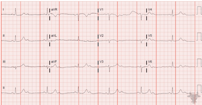
Practice ECG

Answer
Rate: ~80
Rhythm: Sinus
Axis: Normal
P waves: upright in II, biphasic in V1, no evidence of left/right atrial enlargement
PR interval: Normal
QRS: > 120 ms; broad S wave in V1 (without visible r wave). Broad notched R wave in I, broad R wave in V6. No Q waves. Does not quite meet LVH criteria.
ST/T changes: Appropriately discordant STE in V1 (~2mm)
Q waves: not visualized
QT: <500ms
Impression: LBBB


